Endospore Staining Technique- Principle, Requirements, Procedure & Results
Hey, Good to see you here 😀 …… In this Article, we’re gonna discuss Endospore Staining Protocol….. If you have any queries, don’t forget to mention in Comments…
Introduction to Endospore Staining
Some bacteria are capable of changing into dormant structures that are metabolically inactive and do not grow or reproduce. Since these structures are formed inside the cells, hence called endospores. These are remarkably resistant to heat, radiation, chemicals and other agents, that are typically lethal to the organisms. The heat resistance of the spores has been linked to their high content of calcium and dipicolinic acid.
A bacterium forms a single spore by a process called Sporulation. Sporulation takes place either by depletion of an essential nutrient or unfavorable environmental conditions. During Sporulation, vegetative cells give rise to a new intracellular structure termed as “Endospores” that is surrounded by impermeable layers called spore.
The position of the spores may vary from species to species. The spores may be located in the middle of the cell, at the end of the cell (Terminal spores), or between the end and middle (Sub terminal spores) of the cell. Also, Spores may vary in the shape it may be spherical or elliptical.
What is the Principle / Mechanism of Endospore Staining ?
The method of endospores staining was first proposed by Dorner in 1922 which later modified by Schaeffer and Fulton in 1933 which makes the process much easier and effective.
The spores are differentially stained by using the special procedures that help the dyes to penetrate more effectively in the spore wall and stains it. An aqueous solution of primary stain (Malachite Green) is applied and steamed to enhance the penetration of the dye in impermeable spore coat.
Once stained the endospores readily decolorize by using the Tap water/ Distilled water as the Malachite green is water soluble and the wall of the bacterial spore becomes so much weak due to heating that it cannot retains the primary stain and washing with water is enough to decolorize the cell. However, there is no effect observed on the bacterial spore and it appears as green color body inside the cell. Finally, the slide is counterstained with safranine which stains the bacterial cell and the result observed as the green color spores within the red/pink color cells.
The Materials Required For Endospore Staining Protocol
- Glass Slides
- Specimen/Bacterial culture
- Tissue paper
- Inoculating loop
- Spirit lamp/Bunsen burner
- Staining tray
- Microscope
- Wash Bottle
- Tap water/ Distilled water
- Safranine (2.5% aqueous solution)
- Malachite green (5% aqueous solution)
The 0.5% aqueous solution of Malachite green is prepared as follows:
- 5 gm of malachite green.
- 100 ml of distilled water
Mix the 0.5 gm of Malachite green powder in the 100 ml of Distilled water.
Prepare the 2.5% aqueous solution of safranine as follows:
- 5 gm of safranine O
- 100 ml of 95% ethanol
Mix the 2.5 gm of Safranine O powder in 100 ml of 95% Ethanol.
Here is the Procedure Of Endospore Staining
[wp-svg-icons icon=”point-right” wrap=”i”]Take a clean grease free glass slide and make a smear from the culture or the specimen.
[wp-svg-icons icon=”point-right” wrap=”i”]Air dry and heat fix the organism on a glass slide and cover it with a square of blotting paper of appropriate size.
[wp-svg-icons icon=”point-right” wrap=”i”]Saturate the blotting paper with malachite green stain solution, gently heat over the Bunsen burner and steam it for 5 minutes, keeping the paper moist and adding more dye as required. Alternatively, it can be done by placing the slide over a beaker of boiling water.
[wp-svg-icons icon=”point-right” wrap=”i”] Remove the blotting paper from the slide, and rinse the slide gently with tap water or Distilled water using a wash bottle. Dispose of the used blotting paper in the trash as it may contain some spores.
[wp-svg-icons icon=”point-right” wrap=”i”]Counterstain the smear with5% safranine solution for 30 seconds. Wash with distilled water.
[wp-svg-icons icon=”point-right” wrap=”i”]Dry the slide with absorbent paper or blotting paper.
[wp-svg-icons icon=”point-right” wrap=”i”]Examine the slide under the microscope for the presence of endospores.
The Interpretation of Endospore Staining Results
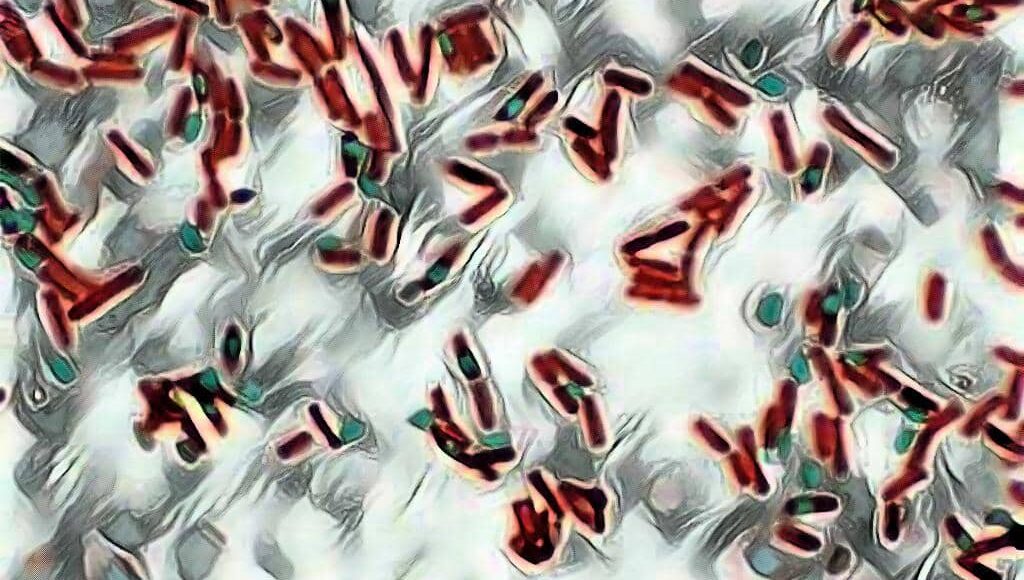
The Cells containing endospores appears as the red colored rod-shaped structure along with an intracellular spherical or elliptical green colored structure. It represents the red colored vegetative bacilli with green colored endospores (intracellular spores).
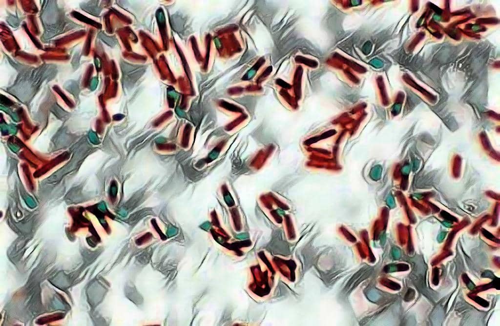
The non-sporing bacterial cell appears red in color but no intracellular green color structures present which makes it easier to distinguish the sporing & non-sporing cells.