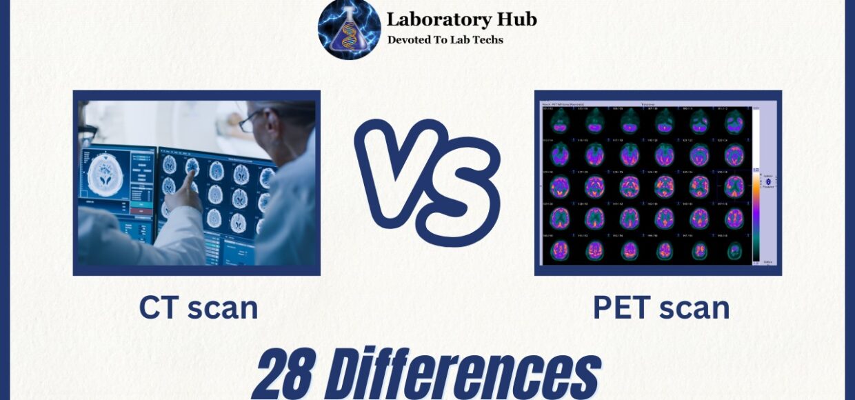
28 differences between CT scan and PET Scan
Welcome to our blog section where we will explore the fascinating world of medical imaging techniques! Today, we delve into the intriguing differences between two commonly used scans: CT scan and PET Scan. These advanced diagnostic tools have revolutionized medical practices, enabling doctors to gain valuable insights into the human body like never before. So, let’s embark on this journey together as we uncover their benefits and side effects!
Both CT scans and PET Scans offer unique advantages in diagnosing various conditions. A CT scan (Computerized Tomography) provides detailed images by combining multiple X-ray views from different angles, allowing doctors to visualize bones, organs, and tissues in remarkable clarity. It is especially effective for detecting fractures, tumors, infections, or bleeding. On the other hand, a PET Scan (Positron Emission Tomography) offers dynamic information about metabolic activity within organs at a cellular level. By injecting a small amount of radioactive material into the body which emits positrons (positively charged particles), it creates three-dimensional images displaying areas with abnormal functioning cells or potential cancerous growths.
While both scans are generally safe procedures performed under professional supervision, they do come with some potential side effects worth considering. CT Scans expose patients to minimal radiation levels that are usually well-tolerated but may be of concern for pregnant women or individuals who require frequent examinations due to cumulative exposure risks. PET Scans involve radiopharmaceutical injections that can cause mild allergic reactions such as itching or rashes.
Also Read: Introns vs Exons- 25 Major Differences
S.No | Aspect | CT Scan | PET Scan |
1 | Full form | Computed Tomography | Positron Emission Tomography |
2 | Imaging modality | X-rays | Radioactive tracers |
3 | Type of images produced | Cross-sectional slices | Metabolic activity maps |
4 | Radiation exposure | Moderate to high | Low |
5 | Use of contrast agents | Commonly used | Occasionally used |
6 | Image resolution | High | Lower than CT |
7 | Anatomical visualization | Excellent | Limited |
8 | Functional visualization | Limited | Excellent |
9 | Applications | Visualize bone and soft tissue structures | Detect cancer, heart problems, brain disorders |
10 | Cancer detection | Limited | Excellent |
11 | Neurological disorders | Limited | Excellent |
12 | Radiation therapy planning | Commonly used | Occasionally used |
13 | Brain imaging | Less effective | More effective |
14 | Alzheimer’s disease detection | Limited | Useful in early detection |
15 | Cardiovascular disease imaging | Limited | Excellent |
16 | Spatial resolution | Higher | Lower |
17 | Time required for the scan | Quick | Longer |
18 | Attenuation correction | Not applicable | Essential for accurate results |
19 | Fusion with other imaging | Commonly done | Commonly done |
20 | Anatomical landmarks | Helpful in identifying | Not clearly defined |
21 | Evaluation of bone fractures | Excellent | Limited |
22 | Evaluation of soft tissue | Limited | Excellent |
23 | Image artifacts | Less likely | Possible due to biological processes |
24 | Cost | Relatively lower | Relatively higher |
25 | Patient preparation | Simple | May require fasting |
26 | Whole-body imaging | Possible | Commonly used |
27 | Image interpretation | Dependent on radiologist’s expertise | Requires specialized training |
28 | Follow-up imaging | Commonly used | Commonly used |
Also Read: Diarrhea vs Dysentery- Definition and 25 Major Differences
Frequently Asked Questions (FAQS)
A CT Scan (Computed Tomography) uses X-rays to produce cross-sectional images of the body, providing detailed anatomical information. On the other hand, a PET Scan (Positron Emission Tomography) utilizes radioactive tracers to visualize metabolic activity, offering functional insights into tissues and organs.
PET Scan is more effective for cancer detection due to its ability to identify abnormal metabolic processes in tissues, allowing for early detection and precise staging of cancer.
CT Scans do involve moderate to high radiation exposure. However, the benefits of accurate diagnosis and treatment planning often outweigh the potential risks, especially when used judiciously by healthcare professionals.
PET Scan can detect changes in brain metabolism and function, which are early indicators of neurological disorders like Alzheimer’s disease. It aids in early diagnosis, disease progression monitoring, and assessment of treatment efficacy.
Yes, CT and PET scans can be fused together to provide a comprehensive evaluation of both anatomical structures and metabolic activity. This combined approach is particularly valuable in oncology and treatment planning.
Yes, PET Scan tends to be relatively more expensive than CT Scan, primarily due to the cost of producing and handling radioactive tracers.
Yes, some PET Scans may require fasting for a certain period before the procedure, as it can affect the distribution of the radioactive tracer in the body.
While CT Scan is commonly used for whole-body imaging, PET Scan is also frequently employed for comprehensive assessments, especially in cancer staging and monitoring disease progression.

