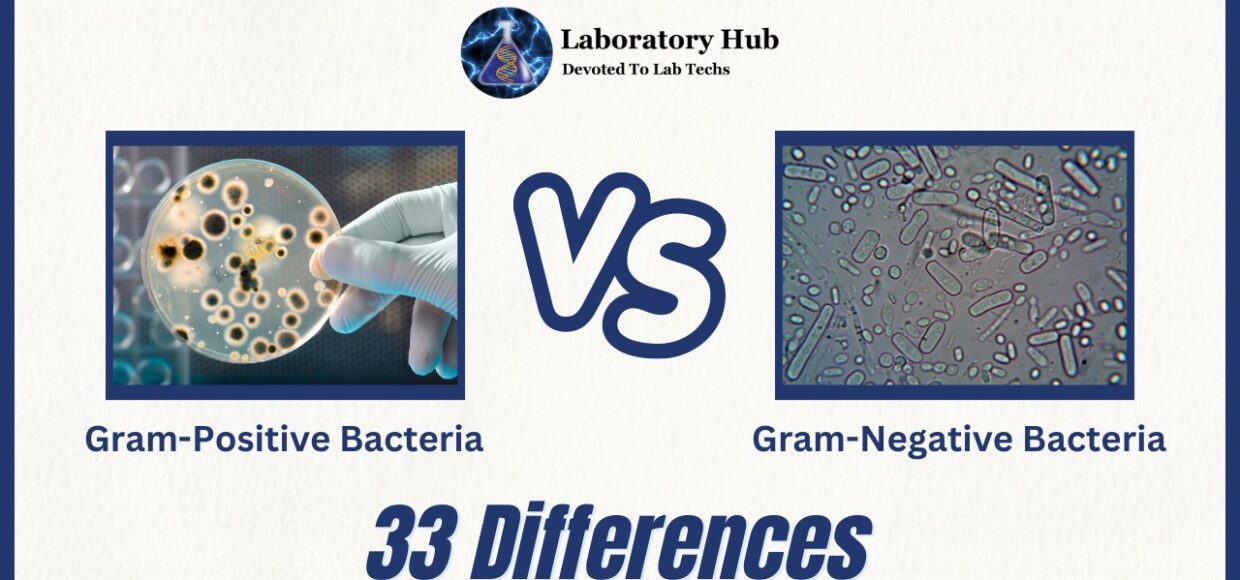
Gram-Positive vs Gram-Negative Bacteria- 33 Differences
Bacteria, the most common and diverse creatures on Earth, impact ecosystems both positively and negatively. A Gram stain-based categorization of bacterial species has revealed their structural and functional traits. Gram-positive and Gram-negative bacteria are classified here. Understanding their behavior, pathogenicity, and antibiotic response requires understanding the major distinctions between these two groups.
The Gram stain technique, created by Danish scientist Hans Christian Gram in the late 19th century, revolutionized microbiology by quickly and accurately distinguishing microorganisms. Bacterial cells are stained with crystal violet dye, iodine, alcohol decolorization, and safranin counterstaining. Based on cell wall composition and structure, the stain’s retention or disappearance classifies bacteria as Gram-positive or Gram-negative.
Gram-positive bacteria’s thick peptidoglycan coating preserves crystal violet stains. This layer shields and supports the bacterial cell. Gram-positive bacteria may also have teichoic acids, lipoteichoic acids, or polysaccharides, which increase antigenicity and host tissue adhesion. Penicillin targets the peptidoglycan layer, making these bacteria more vulnerable to it.
Gram-negative bacteria have a weaker peptidoglycan layer and an outer membrane that does not retain a crystal violet stain. LPS, phospholipids, and proteins in this outer membrane protect against host defenses and make them pathogenic. Porins help Gram-negative bacteria absorb nutrients and expel toxins. The outer membrane also protects Gram-negative bacteria from drugs and the immune system, making them more resistant.
Gram-positive and Gram-negative bacteria have different biological and clinical traits due to structural differences. Gram-positive bacteria cause skin, soft tissue, and respiratory infections. Staphylococcus aureus, Streptococcus pneumoniae, and Enterococcus faecalis. Gram-negative bacteria cause urinary, gastrointestinal, and respiratory illnesses. Escherichia coli, Klebsiella pneumoniae, and Pseudomonas aeruginosa are Gram-negative bacteria.
Clinically, Gram stain categorization helps choose medications and identify diseases. Clinicians can improve patient care by distinguishing Gram-positive and Gram-negative microorganisms.
In conclusion, Gram stain categorization of bacteria has greatly improved our understanding of their structure, behavior, and clinical importance. The different cell wall composition and architecture of these two groups affect their pathogenicity, antibiotic susceptibility, and ecological functions. By studying Gram-positive and Gram-negative bacteria, we can better understand microbiology and fight bacterial illnesses.
Also read: Agglutination vs Precipitation- 20 Differences
S.No. | Category | Gram-Positive Bacteria | Gram-Negative Bacteria |
1 | Cell Wall | Have a thick peptidoglycan layer in the cell wall. | Have a thin peptidoglycan layer in the cell wall. |
2 | Gram Staining | Retain the crystal violet stain and appear purple/blue in Gram staining. | Do not retain the crystal violet stain and appear pink/red in Gram staining. |
3 | Outer Membrane | Lack an outer membrane outside the cell wall. | Have an outer membrane outside the cell wall. |
4 | Periplasmic Space | Have a small or absent periplasmic space between the cell wall and cell membrane. | Have a large periplasmic space between the outer membrane and cell membrane. |
5 | Lipopolysaccharides | Lacks lipopolysaccharides (LPS) in the cell wall. | Contains lipopolysaccharides (LPS) in the outer membrane. |
6 | Teichoic Acids | Some Gram-positive bacteria have teichoic acids in their cell wall. | Lacks teichoic acids in the cell wall. |
7 | Flagella Presence | Can have peritrichous or polar flagella for motility. | Can have peritrichous or polar flagella for motility. |
8 | Flagella Structure | Flagella are composed of two proteins: flagellin and hook. | Flagella are composed of multiple proteins, including flagellin. |
9 | Capsule Presence | Some Gram-positive bacteria have a capsule around the cell wall. | Some Gram-negative bacteria have a capsule outside the cell wall. |
10 | Endotoxins | Lack endotoxins in the cell wall. | Contain endotoxins (lipopolysaccharides) in the outer membrane. |
11 | Exotoxins | Produce exotoxins that are secreted outside the bacterial cell. | Produce exotoxins that are secreted outside the bacterial cell. |
12 | Antibiotic Susceptibility | Generally more susceptible to antibiotics due to a lack of outer membrane. | Can develop resistance to antibiotics due to the presence of efflux pumps and porins. |
13 | Resistance to Drying | More resistant to drying due to the thick peptidoglycan layer. | Less resistant to drying due to the thin peptidoglycan layer and outer membrane. |
14 | Toxicity | Generally less toxic compared to Gram-negative bacteria. | Can be more toxic due to the release of endotoxins. |
15 | Susceptibility to Lysozyme | More susceptible to lysozyme due to the exposed peptidoglycan layer. | Less susceptible to lysozyme due to the outer membrane protecting the peptidoglycan layer. |
16 | Shape | Can have various shapes, including cocci and rods. | Can have various shapes, including cocci, rods, and spirals. |
17 | Efflux Pumps | Generally have fewer efflux pumps. | Can possess multiple efflux pumps, contributing to antibiotic resistance. |
18 | Porins | Lack porins in the outer membrane. | Have porins in the outer membrane, regulating the entry of molecules. |
19 | Antibiotic Targets | Antibiotics target the peptidoglycan layer, cell membrane, or specific intracellular processes. | Antibiotics target the outer membrane, cell wall, or specific intracellular processes. |
20 | Cell Wall Disruption | More susceptible to cell wall-disrupting agents like lysozyme and penicillin. | Less susceptible to cell wall-disrupting agents due to the presence of an outer membrane. |
21 | Electron Transport Chain | The electron transport chain is located in the cell membrane. | The electron transport chain is located in the cell membrane and outer membrane. |
22 | Respiration | Can be facultative anaerobes or obligate anaerobes. | Can be facultative anaerobes, obligate aerobes, or obligate anaerobes. |
23 | Cell Wall Composition | Cell wall primarily composed of peptidoglycan and lipoteichoic acids. | Cell wall composed of a thin peptidoglycan layer and outer membrane containing lipopolysaccharides. |
24 | Outer Membrane Composition | Lacks an outer membrane. | Outer membrane composed of lipopolysaccharides, proteins, and phospholipids. |
25 | Porin Function | Lacks porins in the cell membrane. | Porins in the outer membrane allow the passage of small molecules. |
26 | Membrane Potential | Generates a lower membrane potential. | Generates a higher membrane potential. |
27 | Porin Selectivity | Lacks specific selectivity in porins. | Porins exhibit selective permeability to different molecules. |
28 | Resistance to Detergents | More susceptible to detergents due to the absence of an outer membrane. | More resistant to detergents due to the presence of an outer membrane. |
29 | Cell Envelope Thickness | Generally have a thicker cell envelope. | Generally have a thinner cell envelope. |
30 | Virulence Factors | Produce various virulence factors, including toxins and enzymes. | Produce various virulence factors, including toxins and outer membrane proteins. |
31 | Biofilm Formation | Can form biofilms on surfaces. | Can form biofilms on surfaces. |
32 | Membrane Composition | Cell membrane contains hopanoids, sterols, or glycolipids. | Cell membrane contains hopanoids or sterols but lacks glycolipids. |
33 | Genetic Complexity | Generally have a simpler genetic structure. | Generally have a more complex genetic structure. |
Frequently Asked Questions (FAQs)
Gram-positive bacteria retain crystal violet stains due to their strong peptidoglycan cell walls. Gram-negative bacteria have a thinner peptidoglycan layer and an outer membrane that does not stain.
Antibiotic susceptibility depends on anatomical differences between the groups. Penicillin, which targets the thick peptidoglycan coating of Gram-positive bacteria, is more effective. Gram-negative bacteria, with their extra outer membrane, are more resistant to antibiotics.
Gram-positive and Gram-negative bacteria may infect people and animals. Each category has microorganisms that cause certain ailments. Gram-positive bacteria cause skin, soft tissue, and respiratory infections, whereas Gram-negative bacteria produce urinary tract, gastrointestinal, and respiratory infections.
Gram stain is used in clinical settings to quickly identify germs. The staining pattern helps doctors choose medications and treatment plans for Gram-positive and Gram-negative bacteria.
Bacteria are classified by cell wall construction into Gram-positive and Gram-negative categories to help understand their ecological activities. Gram-positive bacteria in soil, water, and the human microbiome help in nutrient cycling and fermentation. Gram-negative bacteria are abundant in many ecosystems and interact with plants, animals, and other microbes to fix nitrogen and decompose.

