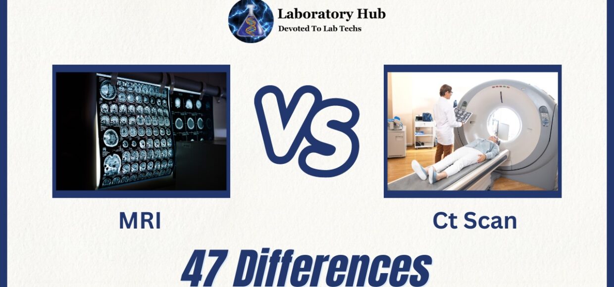
47 Differences Between MRI and CT scan
In the realm of medical imaging, Magnetic Resonance Imaging (MRI) and Computed Tomography (CT) scans are two advanced diagnostic tools that have transformed the way medical professionals visualize and understand the human body. Both techniques are pivotal in diagnosing a wide range of conditions, yet they operate on distinct principles, offer different types of information, and are applied based on the specific clinical context. Understanding the differences between MRI and CT scans is crucial for healthcare providers to make informed decisions about which modality to use for a particular patient and condition.
MRI
MRI, a non-invasive imaging technique, harnesses the power of strong magnetic fields and radiofrequency pulses to create detailed, cross-sectional images of the body’s internal structures. These images offer exquisite soft tissue contrast, making MRI especially valuable for visualizing soft organs like the brain, spinal cord, muscles, and joints. The absence of ionizing radiation in MRI ensures its safety for repeated use, even in delicate situations like monitoring fetal development during pregnancy. However, its longer scan times and potential for claustrophobia pose challenges for some patients.
Also Check: 28 differences between CT scan and PET Scan
CT scan
In contrast, CT scans utilize X-ray technology to create detailed cross-sectional images of the body. A rotating X-ray source and detectors generate multiple images, which are then reconstructed into three-dimensional representations. CT scans excel in capturing anatomical details, making them an ideal choice for diagnosing bone fractures, detecting tumors, and assessing internal bleeding. They are particularly useful in emergency cases due to their speed and comprehensive imaging capabilities. However, the use of ionizing radiation in CT scans demands judicious consideration, especially when multiple scans are required.
Differences Between MRI and CT scans
The differences between MRI and CT scans extend beyond their underlying principles. MRI excels in visualizing soft tissues with exceptional detail, making it invaluable for differentiating between normal and abnormal structures. It is also the preferred choice for evaluating neurological and musculoskeletal conditions where soft tissue information is crucial. On the other hand, CT scans offer quicker imaging times, making them better suited for unstable patients and emergency situations. Additionally, they are more effective in capturing bone and lung images due to their superior contrast.
In conclusion, while both MRI and CT scans are indispensable tools in modern medical practice, they cater to distinct diagnostic needs. MRI prioritizes superb soft tissue contrast, safety, and the absence of ionizing radiation, making it ideal for assessing intricate structures. CT scans excel in capturing anatomical details and speed, rendering them an excellent choice for emergency cases and conditions involving bones or the chest. By comprehending the strengths and differences of MRI and CT scans, healthcare providers can ensure accurate diagnoses and optimal patient care.
Also Check: 38 Differences Between DHA And EPA
Detailed Comparison MRI vs CT scan
S.No. | Aspect | MRI (Magnetic Resonance Imaging) | CT Scan (Computed Tomography) |
1 | Principle | Utilizes strong magnetic fields and radio waves to generate images. | Uses X-rays to create cross-sectional images of the body. |
2 | Radiation Exposure | No ionizing radiation exposure, making it safer for patients. | Involves exposure to ionizing radiation due to X-rays. |
3 | Contrast Enhancement | Uses contrast agents for enhanced visualization of certain tissues. | Can use contrast agents for enhanced visualization of certain tissues. |
4 | Soft Tissue Differentiation | Offers exceptional soft tissue differentiation due to its principle. | Offers good soft tissue differentiation but not as precise as MRI. |
5 | Bone Visualization | Less effective for visualizing bones compared to CT. | Excellent for visualizing bones and dense tissues. |
6 | Internal Bleeding Detection | Effective in detecting internal bleeding and injuries. | Can detect internal bleeding and injuries effectively. |
7 | Brain Imaging | Offers detailed brain imaging and detects abnormalities effectively. | Useful for brain imaging but may have limitations in certain cases. |
8 | Blood Vessel Imaging | Provides detailed images of blood vessels with contrast agents. | Provides detailed images of blood vessels using contrast agents. |
9 | Organ Visualization | Offers detailed visualization of organs and structures. | Provides detailed visualization of organs and structures. |
10 | 3D Reconstruction | Allows 3D reconstruction of images for detailed analysis. | 3D reconstruction is possible for detailed analysis. |
11 | Bone Fractures | Not the first choice for detecting bone fractures. | Good for detecting and visualizing bone fractures. |
12 | Cancer Detection | Effective for detecting tumors and cancerous growths. | Effective for detecting tumors and cancerous growths. |
13 | Abdominal Imaging | Offers detailed images of abdominal organs and structures. | Provides detailed images of abdominal organs and structures. |
14 | Radiation Dose | No ionizing radiation, making it safer for frequent use. | Involves higher radiation dose compared to MRI. |
15 | Pregnancy Imaging | Safe for pregnancy imaging due to no radiation exposure. | Limited use during pregnancy due to radiation exposure. |
16 | Kidney Stone Detection | Limited ability to visualize small kidney stones. | Effective in detecting kidney stones and urinary tract issues. |
17 | Musculoskeletal Imaging | Not commonly used for musculoskeletal imaging. | Useful for visualizing bones and joint-related issues. |
18 | Time for Image Acquisition | Requires more time for image acquisition compared to CT. | Requires a few seconds to a few minutes for image acquisition. |
19 | Cost | Generally more expensive due to equipment and technology. | Less expensive due to lower equipment and maintenance costs. |
20 | Metallic Implants | May cause artifacts or distortions around metallic implants. | Generally unaffected by metallic implants. |
21 | Painless Procedure | Painless procedure, no exposure to X-rays or ionizing radiation. | Painless procedure, but patients may need to hold their breath. |
22 | Children Imaging | Used for pediatric imaging due to its safety. | Preferred for children when high-quality images are needed. |
23 | Emergency Use | Can be used in emergencies due to detailed imaging capabilities. | Useful in emergency cases due to quick image acquisition. |
24 | Liver Imaging | Limited resolution for liver imaging compared to CT. | Provides detailed images of the liver and its abnormalities. |
25 | Guidance for Procedures | Offers real-time guidance for procedures like biopsies. | Offers guidance for biopsies and other medical procedures. |
26 | Allergy Concerns | Generally safer, as no contrast agents are used. | Contrast agents may cause allergic reactions in some patients. |
27 | Spatial Resolution | High spatial resolution due to its principle. | High spatial resolution due to cross-sectional imaging. |
28 | Claustrophobia | Can be problematic for claustrophobic patients due to machine design. | Less likely to trigger claustrophobia. |
29 | Limited Soft Tissue Detail | Limited in certain cases due to its principle. | Limited soft tissue differentiation in certain cases. |
30 | Lung Imaging | Limited use for lung imaging due to breathing motion artifacts. | Limited use for lung imaging due to motion artifacts. |
31 | Screening for Diseases | Limited use for disease screening due to longer procedure times. | Effective for detecting and screening certain diseases. |
32 | Specificity | Specificity varies depending on the application. | High specificity for identifying certain conditions. |
33 | Cardiac Imaging | Limited use for cardiac imaging due to motion artifacts. | Useful for cardiac imaging, particularly for coronary assessment. |
34 | Breast Imaging | Used for breast imaging, providing excellent soft tissue visualization. | Less commonly used for breast imaging. |
35 | Weight Limitations | Doesn’t have significant weight limitations. | Generally has weight limitations for patient comfort. |
36 | Real-time Imaging | Offers real-time imaging during the procedure. | Real-time imaging during the procedure, offering dynamic information. |
37 | Preparation Required | Typically doesn’t require special preparation. | Sometimes requires preparation like fasting or contrast ingestion. |
38 | Bone Density Measurement | Not commonly used for bone density measurement. | Not commonly used for bone density measurement. |
39 | Tissue Penetration | Limited tissue penetration, better for surface imaging. | Good penetration through tissues, suitable for deep imaging. |
40 | Equipment Size | MRI machines are larger and more complex in design. | CT machines are generally smaller and more accessible. |
41 | Localization Accuracy | Localization accuracy can vary based on operator skill. | High localization accuracy for identifying lesion locations. |
42 | Soft Tissue Variations | Less effective in differentiating certain soft tissues. | Can visualize soft tissue variations effectively. |
43 | Accessibility | Equipment is widely accessible but may have space limitations. | Requires specialized equipment and facilities. |
44 | Operator Skill Dependence | Operator skill plays a role in image quality. | Less dependent on operator skill due to automated imaging. |
45 | Visualization of Blood Flow | Excellent visualization of blood flow using Doppler techniques. | Limited visualization of blood flow compared to MRI. |
46 | Cost-Effectiveness | Generally cost-effective due to no radiation and lower equipment costs. | Often less cost-effective due to radiation exposure and equipment. |
47 | Limitations in Obese Patients | Generally less limited in obese patients due to the absence of radiation. | Can be limited in obese patients due to equipment design. |
Frequently Asked Questions (FAQs)
Soft structures like the brain, spinal cord, muscles, and organs can be seen using MRIs. For the diagnosis of ailments including neurological illnesses, joint problems, malignancies, and injuries, they are particularly helpful.
A contrast MRI makes use of a contrast dye to make specific structures or abnormalities more visible. In some circumstances, such as when checking blood arteries or particular tissues, this can provide additional information.
While CT scans offer useful information, ionizing radiation is exposed throughout the procedure. Before prescribing a CT scan, doctors carefully weigh the risks and advantages.
The clinical question, the area being examined, and the patient’s medical history all play a role in deciding between an MRI and a CT scan. CT is frequently utilized for bone and emergency conditions, whereas MRI is excellent for soft tissue detail
That depends on the implant kind. While many contemporary implants are MRI-compatible, some might not be MRI-safe. For patients with metal implants that are not compatible with MRIs, CT scans are typically safer.
MRIs are typically regarded as safe during pregnancy, particularly if they are required for medical reasons. Ionizing radiation-containing CT scans are typically avoided during pregnancy unless the advantages clearly exceed the hazards.