Hey, Good to see you here 😀 …… In this Article, we’re gonna discuss the various techniques used in microbiology laboratory for the examination of different types of specimens….. If you have any queries, don’t forget to mention in Comments…Thanks
Microbiological laboratory testing plays a key role in the management of infection. Accurate and rapid identification of significant microorganisms is vital for guiding optimal antimicrobial therapy and improving outcome from infections and infectious disease.
Laboratory diagnosis is also essential for effective infection prevention and control in both the hospital and community settings, as well as providing valuable epidemiological data.
In addition, accurate diagnosis reduces the over-prescribing of antimicrobial agents and contributes significantly to reductions in the emergence and spread of antimicrobial resistance.
Specimen collection is the crucial step in the investigation procedure of a pathological condition followed by the proper transportation of the specimen to the laboratory and then the identification procedure. The specimen should be collected according to the site of lesion or infection.
What type of Specimen do you require for what kind of Assay?
Pus Sample – Pus sample should be obtained in case of Pyogenic infection to identify the causative agent of the infection. When collecting the pus specimen obtain as much material as possible which may increase the chance of isolating microorganisms which may be difficult to grow or are minimal in number.
Blood Sample – Blood sampling should be performed by a well-trained healthcare worker, as there are many different microbiological and virological blood tests especially the serological tests, performed for the diagnosis of various diseases. The blood specimen is usually analyzed in case of Bacteremia or septicemia or when the blood culture is required.
Cerebrospinal Fluid – Sampling of cerebrospinal fluid (CSF) is essential for the accurate diagnosis of infectious meningitis and may aid in the diagnosis of encephalitis. Cerebrospinal fluid is most commonly obtained via a lumbar puncture performed by medical staff. A sterile technique is required as there is a risk of introducing infection itself causing meningitis. Specimens of CSF should be dispatched to the laboratory immediately.
Ear Swabs – The ear swabs are frequently obtained for the detection of microbes associated with the pathology of the ear. The secretions or exudates from the ear are collected in case of suppurative lesions.
Eye swabs or scrapings – Conjunctival scraping should be collected in case of eye infections especially in the case of Conjunctivitis.
Hair, Nail or Skin – Hair, Nail of Skin specimen can be collected in case of infections of the respective sites.
Nose Swabs – commonly used for the screening of MRSA in the nasal cavity and other microbes that causes the upper respiratory tract infections like Streptococcus pneumoniae.
Sputum Sample – Sputum samples are essential for the accurate microbiological diagnosis of pneumonia, tuberculosis, acute tracheitis, and bronchitis etc..
Stool Sample – Stool specimen is frequently collected to identify parasites and microorganisms that cause diarrhea and bowel disorders.
Throat Swabs – Frequently collected to identify the microbes causing pharyngitis or upper respiratory tract infections.
Urine Sample – Urine samples sent to the microbiology laboratory are for bacteriological investigation associated with Urinary tract infections.
Vaginal/Vulval Swabs – In microbiology, Vaginal swabs are collected for the examination of the vaginal section for the suspected infections.
Wound Swabs – wound swabs are collected for the examination of the secretions or exudates in case of suspected infection.
Some important Guidelines for the Specimen Collection
All the samples should be collected in specific sterile containers by taking all the necessary precautions and under strict aseptic conditions to prevent the contamination.
Sterile Cotton swabs should be used for the collection of the specimens from the sites such as Nose or Perineum and immediately the swabs are transferred to the test tubes containing Normal saline or Peptone water or directly inoculated onto the sterile media plates.
In case of the Urine specimen, Mid-stream urine should be collected to prevent the contamination of the Normal flora or other microbes that may be present on or near to the urethral opening.
The blood specimen should be collected in the appropriate culture bottles as per the age of the patient (Children or Adult) and it should contain the Glucose broth &/or Taurocholate broth.
In case of delicates organism, special transportation medium should be used that are specific for the microorganism.
The Transportation of Specimen to Microbiology Laboratory
All specimens must be appropriately labeled with the patient’s name and identification number assigned or the barcode slip should be properly labeled on the container before transporting to the microbiology laboratory.
All the specimens should be transported & processed immediately as soon as the specimen reaches the laboratory to prevent the erroneous results.
Macroscopic Examination of the Specimen in Microbiology Laboratory
The macroscopic examination of the specimen includes the physical & chemical examination of the specimen. The physical examination includes the analysis of the specimen for the Color, Consistency, Odour, Specific gravity, pH, Volume, and Appearance Whereas the Chemical Examination of the specimen includes the Detection of Glucose, Protein, Bilirubin & Ketone bodies in the specimens directly and various biochemical tests are performed in the microbiology laboratory from the specimens directly or from the colonies obtained by culturing the specimen on proper culture media.
Like in the pus specimen, the consistency of the pus is examined which varies from the thin clear watery fluid to the Thick milky fluid. The color of the pus depends on the type & severity of infection, which may be white, White-yellow, Yellowish-Brown or Green.
In Urine specimen, Beside the Culture, Antibiotic sensitivity test & microscopic examinations; Volume, color, Specific gravity, pH, and Appearance is checked in the physical examination as well as the Glucose, Protein, Bilirubin, Bile salts, Ketone bodies, and Occult blood are detected as Chemical examination.
However, the Physical and chemical examination of urine are the parts of the Clinical Pathology laboratory and the Culture & Antibiotic sensitivity is done in the Microbiology laboratory.
In Microbiology laboratory, the Cerebrospinal fluid (CSF) is Examined for the detection of any microorganism, especially in case of Meningitis and Myelitis.
In the Physical examination of the CSF, the Color, Viscosity, Pressure, and Turbidity is checked whereas in chemical examination glucose and Protein are analyzed and sometimes the IgG, LDH, CRP, and Protein electrophoresis is done which is mainly performed in the Pathology and Biochemistry departments.
The Sputum specimen is examined for the color and consistency which reveals many pathologies of the Respiratory tract. Especially the sputum examination is prescribed in case of suspected tuberculosis and sometimes in pneumonia.
The Culture and microscopic examination, as well as certain biochemical tests, can be done to confirms the detect the presence of the microorganism.
Differential and Specific staining techniques like Gram staining and Acid fast stainings are done on the smears prepared directly from the specimen or from the colonies obtained from the culture of the specimen.
Macroscopically, the stool samples are examined for the color and consistency as for whether the fecal matter is semisolid or fluid and whether or not it contains the Blood, Mucus, pus and the segments of some parasites like Tapeworms and Roundworm.
The color of stool also indicates the various pathologies that may be present in the GIT like the Green stool are present in case of infantile diarrhea, Clay-colored in case of obstructive jaundice, excess fats etc. Dark brown or Bright red in case of Bleeding from distal colon and black is common in GIT bleeding and Intestinal TB. The chemical examination of the stool is mainly done for the pH.
Some bacteria are the normal inhabitant of the GIT tract and are commonly found in the stool samples which includes the E.coli, Klebsiella, Enterobacter and Pseudomonas species.
The microbiological examination is done for the detection of the pathogenic bacteria or parasites that cause the pathologies of the GIT. Commonly, the culture of the stool sample is done for detecting the Shigella, Salmonella and Campylobacter species and the bacteria that commonly associated with diarrhea like Vibrio, Yersinia, Enterocolitica & Aeromonas species.
Microscopic Examination of the Specimen in Microbiology Laboratory
Here comes the Crucial role of microbiology department in analyzing the specimen microscopically for detecting the microorganism that is causing the pathology or disease in the individual’s body….
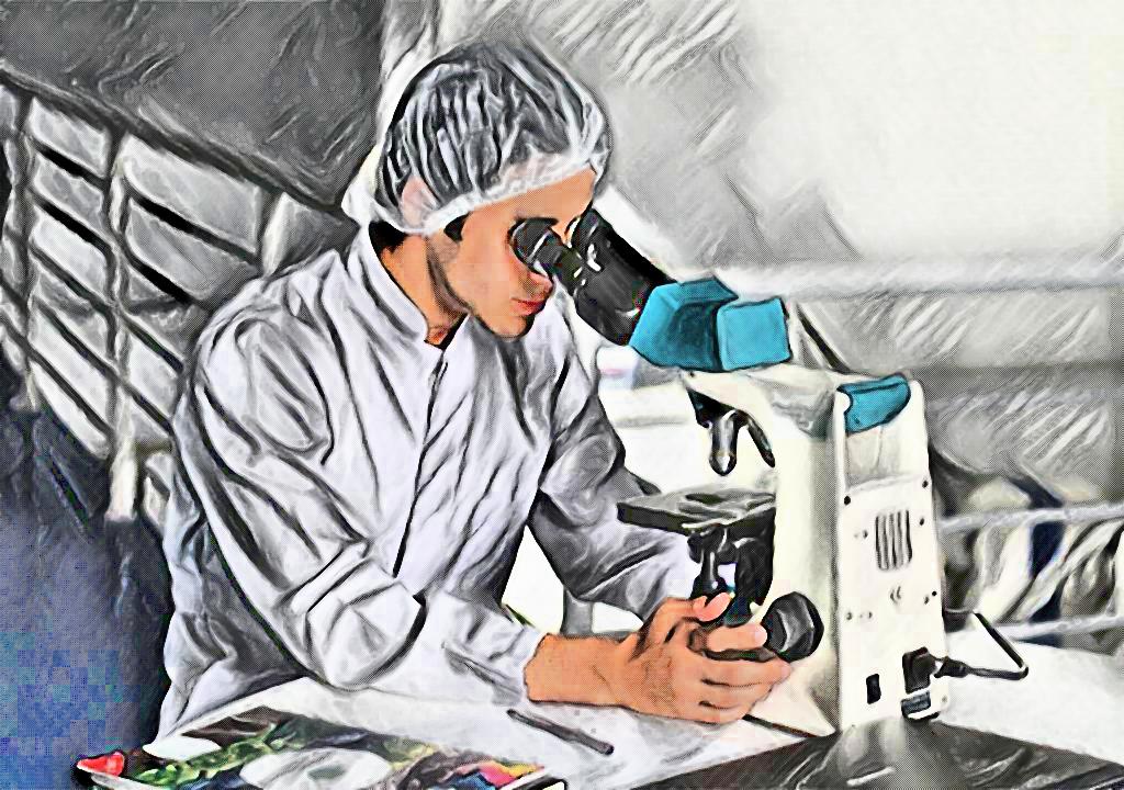
The first step in this is the direct examination of the specimen under the microscope by making a thin smear on the microscopic glass slide and staining it with a differential staining like Gram’s Staining which gives some idea to the pathologist that what kind of a microbe is suspected to be present and the further procedure of the specimen examination is done accordingly.
Afterward, the culture of the specimen is done to isolate the microbe in pure cultures. For this, the specimen is inoculated on the suitable sterile culture media plates and incubated at an optimum temperature in the incubator, commonly at 37°C.
Various types of media are used in the microbiology laboratory for the isolation of microorganisms present in the specimen. Some of the commonly used media are as follows:
- Nutrient Agar Medium
- Blood Agar Medium
- McConkey Agar Medium
- Sabouraud Dextrose Agar
Selective media like Selenite F broth, Salmonella-Shigella agar, and Xylose-Lysine Deoxycholate (XLD) medium are used for the isolation of Salmonella and Shigella species, Thiosulfate Citrate Bile salt agar (TCBS) for Vibrio species, Campylobacter Blood Agar (CAMPY-BA) medium for Campylobacter species, Cycloserine Cefoxitin Fructose agar (CCFA) for Clostridium deficille, Lowenstein Jensen (LJ) medium for Mycobacterium tuberculosis, Mannitol Salt Agar (MSA) for Staphylococcus are also used for the isolation of respective microbes.
The Sabouraud Dextrose agar is the selective media for the growth of Fungal Cultures as it inhibits the growth of most of the bacteria by adding some antibiotics like Tetracycline and Chloramphenicol the bacteria inhibiting property is enhanced and the pure colonies of fungi can easily be obtained.
After inoculation the plates are incubated at the optimum temperature like for most of the bacteria is 37°C and for the fungi is 25°C for about 24 – 48 hours. The fungi are incubated for about 7-10 days as fungi take much longer time to grow on artificial culture media.
After this the culture plates are examined for the colony morphology as the Size, Shape, Color, Structure, Elevation, Surface, Emulsifiability, edges, Consistency etc. which may give a microbiologist a rough idea about the suspected microbe present.
Afterward, the colonies obtained in the culture of the specimen are analyzed for the identification of true pathogen that is causing the pathology.
For this, Initially the microscopy is done by making the smear from the culture obtained and staining it using the suitable staining techniques Like Gram staining which differentiates the gram positive and gram negative bacteria, Acid fast staining which differentiates Acid fast and non-acid-fast bacteria, Albert staining for observing the Metachromatic property, Endospore staining for the detection of Endospores, Negative staining for detecting the presence or absence of capsule and so on…..
The colonies are further analyzed for the biochemical tests like Sugar fermentation, Indole production, MR test, VP test, Citrate utilization, Citrate production, Coagulase production, Nitrate reduction, Urease test, Oxidase reaction and much more which are done according to the microbe suspected which make it much easier to get to the conclusion that which microbe is associated with the pathology.
The Antibiotic Sensitivity testing is done on the colonies obtained from the culture of the specimen to know that which antibiotic(s) are effectively working on the microbe and helps the doctor to prescribe the effective treatment to the patient.
Beside these Culture and Antibiotic testing, Various serological tests are done which are based on Antigen-Antibody reaction especially to detect the viral diseases like HIV infection, Hepatitis B & C infection, CMV infection etc. which is done by detecting the presence of Antigens or Antibodies in the patient’s serum.
Commonly used techniques for this is the Enzyme-Linked ImmunoSorbent assay (ELISA). Qualitative Assay kits are commercially available nowadays which give the results in just a few minutes. as well as the quantitative assays are used to know the severity of the infection, WIDAL qualitative, as well as quantitative test for the diagnosis of Salmonella antigens, is based on the Antigen-Antibody reaction which is most commonly used for the detection of Salmonella infection.
In this way, the specimens are analyzed in the microbiology laboratory for the detection of microbes that are associated with the pathological condition.
Frequently Asked Questions (FAQs)
Q1. How do you collect a specimen sample?
Specimen collection techniques depend on the type of sample being collected. Generally, a sterile collection container is used to collect the sample. The area being sampled is cleaned with an antiseptic to reduce contamination. Examples of collection techniques include swabbing, aspiration, biopsy, and urine collection.
Q2. What is the laboratory method of microbiology?
Microbiology laboratory methods involve the cultivation, isolation, and identification of microorganisms. Microbiologists use a variety of techniques to culture microorganisms on agar plates or in liquid media. These methods include streaking, pouring, and spread plating. Once the microorganisms are grown, they can be identified by morphological, biochemical, and molecular techniques.
Q3. What is the method for microbiological sampling?
Microbiological sampling involves collecting samples from different sources, such as air, water, surfaces, or biological materials. The method used for microbiological sampling depends on the type of sample being collected. For example, air samples may be collected using impaction, filtration, or sedimentation methods. Water samples may be collected using grab or composite sampling techniques. Surface swabs, contact plates, and settle plates may be used for surface sampling.
Q4. How is microbiology test done?
Microbiology tests involve the cultivation, identification, and characterization of microorganisms. Cultivation involves the growth of microorganisms on agar plates or in liquid media. Identification can be done by observing the morphological and biochemical characteristics of the microorganism. Characterization involves testing the microorganism’s susceptibility to antibiotics or its ability to produce specific enzymes or pigments.
Q5. What methods are used to identify bacteria in microbiology?
There are several methods used to identify bacteria in microbiology, including Gram staining, biochemical tests, and molecular methods. Gram staining is a rapid way to distinguish between gram-positive and gram-negative bacteria. Biochemical tests involve the use of specific reagents to test for the presence or absence of enzymes or other molecules. Molecular methods, such as PCR and DNA sequencing, can be used to identify bacteria based on their genetic material.
Q6. What are three methods used to identify microorganisms in the laboratory?
Three methods used to identify microorganisms in the laboratory are morphological, biochemical, and molecular methods. Morphological methods involve observing the physical characteristics of the microorganism, such as shape, size, and motility. Biochemical methods use specific reagents to test for the presence or absence of enzymes or other molecules. Molecular methods, such as PCR and DNA sequencing, can be used to identify microorganisms based on their genetic material.
User Review
( votes)Laboratory Hub aims to provide the Medical Laboratory Protocols & General Medical Information in the most easy to understand language so that the Laboratory Technologist can learn and perform various laboratory tests with ease. If you want any protocol to be published on Laboratory Hub, Please drop a mail at contact@laboratoryhub.com. Happy Learning!