Hey, good to see you here 😀 …… In this article, we’re gonna discuss the Principle, Requirements, Procedure & Results of Albert Staining….. If you have any queries, don’t forget to mention in comments….. Thanks
What is Albert Staining Technique?
Corynebacterium is the genus of Gram +ve, non-acid-fast, non-sporing, non-motile bacilli.
The most important member of the genus is Corynebacterium diphtheriae, the causative agent of Diphtheria disease in children.
The C. diptheriae or diphtheria bacillus was first described by Klebs but Loffler was the first to cultivate it in the laboratory and also called as the Klebs-Loffler’s bacillus (KLB)
They form metachromatic granules, these granules are formed by many other types of bacteria.
They are made up of Polyphosphate and considered as the food reserve in bacteria. These food reserves are involved in all metabolic processes.
In sputum slide, Albert’s staining is used to differentiate the gram +ve bacteria forming metapositive chromatic granules or Volutin granules. These bacteria are present in a CLUB or Chinese letter arrangement.
Principle Of Albert Staining
The bacterial cell of Corynebacterium diphtheriae & Yersinia pestis has volutin granules in their cytoplasm which are highly acidic while the cytoplasm is neutral.
Albert stain I have two dyes ‘Toluidine blue O’ and ‘Malachite green’ both of which are basic dyes with high affinity for neutral tissue components like cytoplasm and the pH of Albert stain I is adjusted to 2.8 by using ‘Glacial acetic acid’, which is acidic for cytoplasm (as it is neutral) but basic for volutin granules (as the pH of volutin granules are highly acidic).
Therefore, When Albert stain I applied to the cell the volutin granules stain by Toluidine blue O while cytoplasm is green by Malachite green.
Due to the metachromatic property of volutin granules when stained with Toluidine blue O dye they appear Red in color.
When Albert stains II i.e. the Iodine solution is applied due to the effect of Iodine the metachromatic property is not observed and Granules appear blue-black in color.
The Requirements For Albert Staining
- Glass Slides
- Specimen/Bacterial culture
- Tissue paper
- Inoculating loop
- Spirit lamp/Bunsen burner
- Staining tray
- Microscope
- Wash Bottle
- Albert’s stain A
- Albert’s Stain B
The Composition Of Albert Stain:
Albert’s A Solution consist of
- Toluidine blue 0.15 gm
- Malachite green 0.20 gm
- Glacial acetic acid 1 ml
- Alcohol (95% ethanol) 2ml
Dissolve the dyes in alcohol and add to the distilled water then add the acetic acid to the thus obtained solution. Allow the stain to stand for one day and then filter. Add Distilled water to make the final volume 100ml.
Albert’s B Solution consists of
- Iodine 2gm
- Potassium iodide (KI) 3 gm
Dissolve the KI in water and then add iodine. Dissolve iodine in thus formed potassium iodide solution.
Here is the Procedure Of Albert Staining Technique
Take an inoculum of bacterial culture or sputum sample with help of inoculating loop.
Prepare a thin smear of the specimen by mixing the cell with a drop of water / Normal saline on the glass slide.
Heat fix the smear onto the slide by gently passing it over the flame.
Flood the smear with Albert’s Stain A solution and wait for 5 min.
Remove the dye and flood it with Albert’s Stain B for 1 min.
Wash the smear with tap water / Distilled water.
Air dry the slide and observe under the microscope at 100X objective.
Interpretation Of The Albert Staining Results
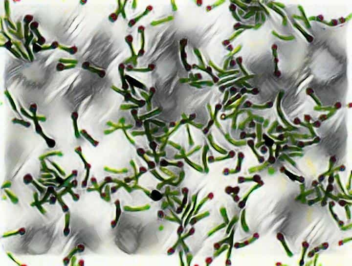
Metachromatic / Volutin granules appeared as a bluish-black in the green colored bacterial body arranged at an angle to each other, resembling English letter ‘L’, ‘V’ or Chinese letter pattern.
 Frequently Asked Questions (FAQs)
Q1. What are the results and interpretation of simple staining?
Simple staining results in the uniform staining of all types of cells or microorganisms. It allows for the visualization of cell shape, size, and arrangement. The interpretation of simple staining is limited to morphological characteristics, as it does not distinguish between different types of microorganisms.
Q2. What is the result and interpretation of Gram staining?
Gram staining results in the differentiation of bacteria into Gram-positive and Gram-negative based on differences in cell wall structure. Gram-positive bacteria stain purple, while Gram-negative bacteria stain pink. The interpretation of Gram staining is valuable for identifying bacterial species and determining appropriate antibiotic treatments.
Q3. What is the result interpretation of acid-fast staining?
Acid-fast staining is used to identify acid-fast bacteria, such as Mycobacterium tuberculosis, which resist decolorization by acid-alcohol after staining with basic dyes. Acid-fast bacteria appear red, while non-acid-fast bacteria appear blue. The interpretation of acid-fast staining is important in the diagnosis of tuberculosis and other related diseases.
Q4. What is the principle of acid-fast bacteria staining?
The principle of acid-fast bacteria staining is based on the unique cell wall structure of acid-fast bacteria, which contains high concentrations of mycolic acid. This allows them to resist decolorization by acid-alcohol, which is used to remove the stain from non-acid-fast bacteria. Basic dyes, such as carbolfuchsin, are used to stain acid-fast bacteria.
Q5. What is the principle of negative staining?
The principle of negative staining is based on the use of acidic stains, such as India ink or nigrosin, which are repelled by the negatively charged bacterial surface. The result is a dark background surrounding the unstained bacterial cells, which allows for the visualization of cell size, shape, and arrangement.
Q6. What reagent is used in acid-fast staining?
Acid-fast staining uses a combination of carbolfuchsin and heat to stain acid-fast bacteria. Acid-alcohol is used as a decolorizing agent, and methylene blue is used as a counterstain to stain non-acid-fast bacteria.
User Review
( votes)Laboratory Hub aims to provide the Medical Laboratory Protocols & General Medical Information in the most easy to understand language so that the Laboratory Technologist can learn and perform various laboratory tests with ease. If you want any protocol to be published on Laboratory Hub, Please drop a mail at contact@laboratoryhub.com. Happy Learning!