Hey, good to see you here 😀 …… In this article, we’re gonna discuss the Principle, Requirements, Procedure & Results of Acid Fast Staining….. If you have any queries, don’t forget to mention in comments….. Thanks
What is Acid Fast Staining technique?
Acid fast staining technique was developed in 1882 by Paul Ehrlich and modified by Ziehl & Nielsen in 1890 hence called Z.N. Staining or Ziehl-Neelsen staining.
Certain bacteria and Actinomycetes have many components in the cell wall and their cell wall has little permeability.
Majority of bacteria can be stained with simple stain but the number of Mycobacteria and Nocardia can only be stained by the special technique known as Acid-fast staining technique.
It was first used for Mycobacterium tuberculosis. Also, this staining technique can be used to stain all the species of Mycobacterium.
What is the Principle Of Acid Fast Staining?
In the cell wall of Acid-fast Bacteria, a lipid called Mycolic acid is present that is a waxy substance.
Mycolic acid resists the staining of the bacterial cell by simple stains or using simple techniques but when Carbol fuchsin & heat is applied, the Mycolic acid layer gets weaken and becomes porous, due to which the carbol fuchsin enters the cell wall.
During decolorization, a special decolorizer containing sulfuric acid or Hydrochloric acid is used.
Bacterial cells that get decolorized and appear colorless were said to be Non Acid- fast bacteria and those which do not get decolorized even with this powerful decolorizer were sad to be Acid-Fast Bacteria.
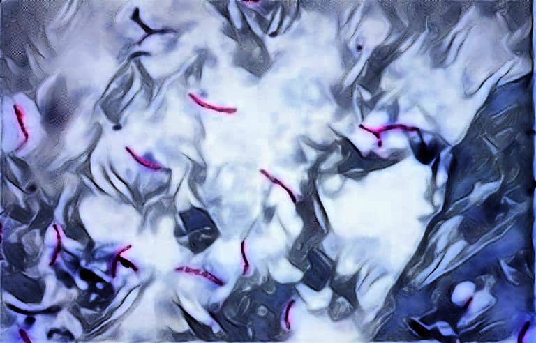
After decolorization, the Acid fast bacteria appear Red in color whereas the Non-acid fast bacteria appear as colorless bodies.
To distinguish them easily a counterstain i.e. Methylene blue is applied which stains the Non-acid fast bacteria and they appear as blue colored bodies under the microscope.
The Requirements For Acid Fast Staining
- Bacterial culture/ Sputum sample
- Glass Slide
- Microscope
- Wash bottle
- Distilled water
- Acid-Fast Decolorizer
- Inoculating loop
- Spirit lamp / Bunsen burner
- Carbol Fuchsin
- Methylene blue
Procedure Of Acid Fast Staining
Prepare a thin smear of the specimen on a clean grease free glass slide.
Fix the smear onto the slide by passing over the flame of Spirit lamp or Bunsen burner.
Gently flood the smear with Carbol Fuchsin dye for 5 minutes.
Gently heats the slide till fumes appear & keep the smear moisten with dye.
Wash the smear with Distilled water using a wash bottle.
Decolorize the smear using Acid-fast decolorizer.
Wash the smear using a wash bottle.
Cover the smear with Methylene blue dye for 1 minute and then wash with distilled water using wash bottle.
Air dry the smear and observe under the microscope at 100X.
Interpretation Of Acid Fast Staining
Acid-fast Bacteria appear Red in color.
Non- Acid fast Bacteria appear blue in color.

check out the interpretation of results & reporting of AFB (acid fast bacilli) in sputum smear
Frequently Asked Questions (FAQs)
Q1. What is the principle of acid-fast bacteria?
Acid-fast bacteria have a waxy layer of mycolic acid in their cell wall, which makes them resistant to many staining techniques. This property is used in identifying them by the acid-fast staining technique, where they retain the stain even after being washed with acid-alcohol.
Q2. What is the principle of acid-fast staining Wikipedia?
Acid-fast staining is a differential staining technique used to identify acid-fast bacteria from non-acid-fast bacteria. It involves using a primary stain, such as carbol fuchsin, which penetrates the waxy layer of the acid-fast bacteria and stains them red. This stain is retained even after treatment with decolorizer, and counterstaining with methylene blue is done to visualize non-acid-fast bacteria.
Q3. What is the principle of acid-fast staining in microbiology?
Acid-fast staining is based on the principle of differential staining, where acid-fast bacteria are differentiated from non-acid-fast bacteria by their ability to retain the primary stain, even after treatment with acid-alcohol. The acid-fast bacteria have a waxy layer that makes them resistant to decolorization, and they appear red under the microscope, while non-acid-fast bacteria appear blue after counterstaining.
Q4. What is the principle of staining procedure?
Staining procedures involve using dyes or stains to visualize biological structures or components under the microscope. The principle of staining procedure is based on the ability of the dye or stain to bind to specific structures or components of the cells, which enhances their contrast and visibility. Different staining procedures are used depending on the structures or components being visualized.
Q5. What is the main principle of staining?
The main principle of staining is to enhance the contrast and visibility of biological structures or components under the microscope by using dyes or stains that bind to specific structures or components. Different staining techniques are used based on the nature of the structures or components being visualized, and the type of microscopy being used.
Q6. What is the principle of staining experiment?
The principle of staining experiment is based on the use of dyes or stains to visualize and identify biological structures or components under the microscope. The experiment involves selecting the appropriate staining technique, preparing the sample, applying the stain, and observing the sample under the microscope to analyze the staining pattern and identify the structures or components of interest.
User Review
( vote)
Laboratory Hub aims to provide the Medical Laboratory Protocols & General Medical Information in the most easy to understand language so that the Laboratory Technologist can learn and perform various laboratory tests with ease. If you want any protocol to be published on Laboratory Hub, Please drop a mail at contact@laboratoryhub.com. Happy Learning!

